Treating an Arteriovenous Malformation (AVM)
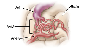
Normally, arteries carry blood with oxygen from the heart to the brain and veins carry blood with less oxygen away from the brain. However, an AVM results in a tangle of blood vessels in the brain. This tangle bypasses brain tissue and sends blood directly from the arteries to the veins. It's important to get medical attention for an AVM as soon as possible. Often, immediate treatment may help prevent serious complications of some AVMs. Current surgical methods make treatment for AVM safer and more effective than ever. The goal of treatment is to stop the flow of blood within the AVM and to prevent it from re-bleeding.

Treatment team
The members of your healthcare team guide you through your treatment, support you, and answer your questions. Your team may include the following specialists:
-
A neurosurgeon. This healthcare provider does the procedure needed to correct your problem.
-
A radiologist. This healthcare provider specializes in using medical imaging in the diagnosis and treatment of disease.
-
A neurologist. This specialist helps co-manage the person during the acute phase of illness, helps direct recovery therapy, and helps manage complications such as seizures.
-
Registered nurses. These professionals provide patient care and teach family and friends to help with your recovery.
-
Physiatrists. These healthcare providers specialize in rehabilitation. They help direct the recovery of lost motor and language skills.
-
Physical and occupational therapists. These providers help with walking, dressing, and other life skills.
-
Speech therapists. These providers focus on speech and swallowing problems.
-
A case manager or social worker. These professionals help guide you through the healthcare system.
Surgery for AVM
Surgical resection removes the tangled blood vessels.
-
Reaching the brain. The surgeon uses a procedure called craniotomy to reach the brain. During a craniotomy, small holes called burr holes are made in the skull. The bone between the holes is cut and lifted away. Finally, the surgeon opens the dura and exposes the brain.
-
Removing the AVM. Once the surgeon has access to the AVM, the abnormal arteries and veins are removed. This redirects blood flow to normal vessels, preventing the AVM from bursting and leaking blood.
-
Closing the skull. When the AVM has been removed, the dura covering the brain is closed. In most cases, the skull bone is put back. The skull bone can be held in place using one of several methods. Titanium clamps are often used, as they provide the most stability and cover the burr holes. After the clamps are in place, the skin incision is closed with stitches or staples.
Other treatments
AVMs sometimes require a combination of treatments. Other treatments include:
-
Radiosurgery. Radiation (beams of gamma rays) can be directed at the AVM using tools called gamma knife, LINAC, or proton beam. The irradiation delivered by the gamma knife closes off the abnormal blood vessels so that blood no longer flows through them. Specialists (neurosurgeon, physicist, and radiation oncologist with special training in this field) will be involved.
-
Embolization. Glue is inserted into the AVM through a catheter (very thin tube). This blocks blood flow in the AVM and holds the blood vessels in a new location. Embolization may happen by itself or as part of the surgical procedure. Beads, tiny balloons, or coils (tiny, spring-shaped objects) can also be used to embolize an AVM. A specialist will be involved in embolization.
© 2000-2024 The StayWell Company, LLC. All rights reserved. This information is not intended as a substitute for professional medical care. Always follow your healthcare professional's instructions.